Terbium »
PDB 6xzf-8u7c »
8bl6 »
Terbium in PDB 8bl6: De Novo Single-Chain Immunoglobulin Dimer SCIG12+EF3A
Protein crystallography data
The structure of De Novo Single-Chain Immunoglobulin Dimer SCIG12+EF3A, PDB code: 8bl6
was solved by
M.Nadal,
J.Roel-Touris,
E.Marcos,
with X-Ray Crystallography technique. A brief refinement statistics is given in the table below:
| Resolution Low / High (Å) | 58.59 / 2.80 |
| Space group | P 43 21 2 |
| Cell size a, b, c (Å), α, β, γ (°) | 73.841, 73.841, 96.245, 90, 90, 90 |
| R / Rfree (%) | 20.2 / 25.7 |
Terbium Binding Sites:
The binding sites of Terbium atom in the De Novo Single-Chain Immunoglobulin Dimer SCIG12+EF3A
(pdb code 8bl6). This binding sites where shown within
5.0 Angstroms radius around Terbium atom.
In total 3 binding sites of Terbium where determined in the De Novo Single-Chain Immunoglobulin Dimer SCIG12+EF3A, PDB code: 8bl6:
Jump to Terbium binding site number: 1; 2; 3;
In total 3 binding sites of Terbium where determined in the De Novo Single-Chain Immunoglobulin Dimer SCIG12+EF3A, PDB code: 8bl6:
Jump to Terbium binding site number: 1; 2; 3;
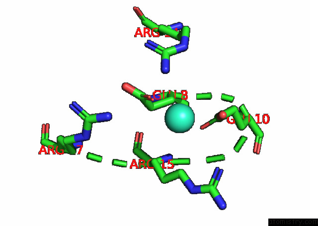
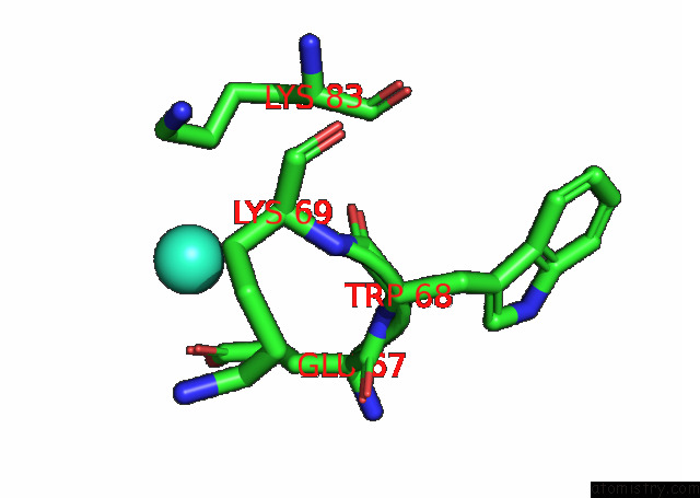
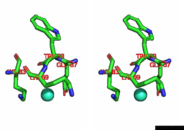
Terbium binding site 1 out of 3 in 8bl6
Go back to
Terbium binding site 1 out
of 3 in the De Novo Single-Chain Immunoglobulin Dimer SCIG12+EF3A
Mono view
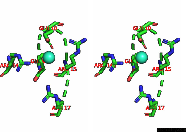
Stereo pair view
Mono view
Stereo pair view
A full contact list of Terbium with other atoms in the Tb binding
site number 1 of De Novo Single-Chain Immunoglobulin Dimer SCIG12+EF3A within 5.0Å range:
|
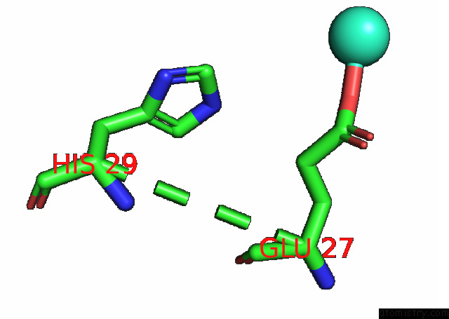
Terbium binding site 2 out of 3 in 8bl6
Go back to
Terbium binding site 2 out
of 3 in the De Novo Single-Chain Immunoglobulin Dimer SCIG12+EF3A
Mono view
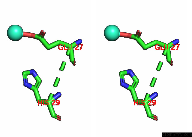
Stereo pair view
Mono view
Stereo pair view
A full contact list of Terbium with other atoms in the Tb binding
site number 2 of De Novo Single-Chain Immunoglobulin Dimer SCIG12+EF3A within 5.0Å range:
|
Terbium binding site 3 out of 3 in 8bl6
Go back to
Terbium binding site 3 out
of 3 in the De Novo Single-Chain Immunoglobulin Dimer SCIG12+EF3A
Mono view
Stereo pair view
Mono view
Stereo pair view
A full contact list of Terbium with other atoms in the Tb binding
site number 3 of De Novo Single-Chain Immunoglobulin Dimer SCIG12+EF3A within 5.0Å range:
|
Reference:
J.Roel-Touris,
M.Nadal,
E.Marcos.
Single-Chain Dimers From De Novo Immunoglobulins As Robust Scaffolds For Multiple Binding Loops. Nat Commun V. 14 5939 2023.
ISSN: ESSN 2041-1723
PubMed: 37741853
DOI: 10.1038/S41467-023-41717-5
Page generated: Fri Oct 11 08:53:58 2024
ISSN: ESSN 2041-1723
PubMed: 37741853
DOI: 10.1038/S41467-023-41717-5
Last articles
Zn in 9MJ5Zn in 9HNW
Zn in 9G0L
Zn in 9FNE
Zn in 9DZN
Zn in 9E0I
Zn in 9D32
Zn in 9DAK
Zn in 8ZXC
Zn in 8ZUF