Terbium »
PDB 1m9s-6xym »
4esq »
Terbium in PDB 4esq: Crystal Structure of the Extracellular Domain of Pknh From Mycobacterium Tuberculosis
Protein crystallography data
The structure of Crystal Structure of the Extracellular Domain of Pknh From Mycobacterium Tuberculosis, PDB code: 4esq
was solved by
A.Cavazos,
D.M.Prigozhin,
T.Alber,
with X-Ray Crystallography technique. A brief refinement statistics is given in the table below:
| Resolution Low / High (Å) | 48.78 / 1.70 |
| Space group | P 1 21 1 |
| Cell size a, b, c (Å), α, β, γ (°) | 47.455, 35.923, 49.308, 90.00, 98.36, 90.00 |
| R / Rfree (%) | 16.3 / 19.7 |
Terbium Binding Sites:
The binding sites of Terbium atom in the Crystal Structure of the Extracellular Domain of Pknh From Mycobacterium Tuberculosis
(pdb code 4esq). This binding sites where shown within
5.0 Angstroms radius around Terbium atom.
In total 2 binding sites of Terbium where determined in the Crystal Structure of the Extracellular Domain of Pknh From Mycobacterium Tuberculosis, PDB code: 4esq:
Jump to Terbium binding site number: 1; 2;
In total 2 binding sites of Terbium where determined in the Crystal Structure of the Extracellular Domain of Pknh From Mycobacterium Tuberculosis, PDB code: 4esq:
Jump to Terbium binding site number: 1; 2;
Terbium binding site 1 out of 2 in 4esq
Go back to
Terbium binding site 1 out
of 2 in the Crystal Structure of the Extracellular Domain of Pknh From Mycobacterium Tuberculosis
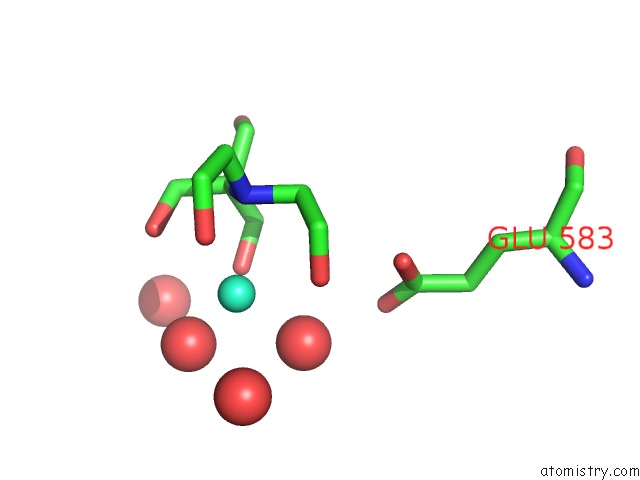
Mono view
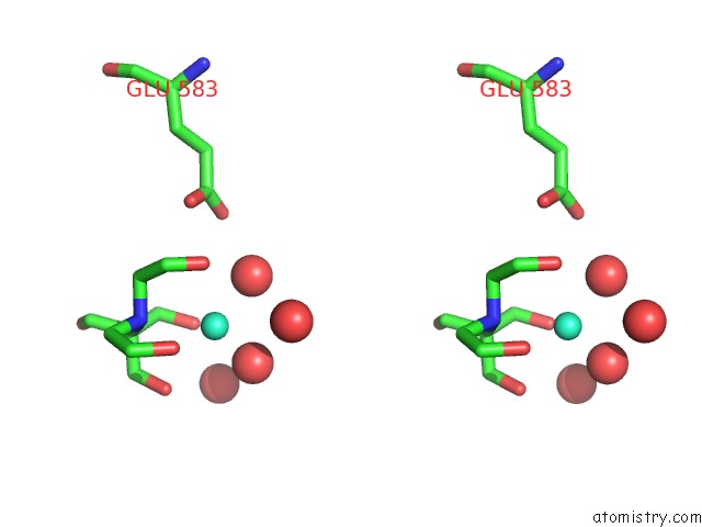
Stereo pair view
Mono view
Stereo pair view
A full contact list of Terbium with other atoms in the Tb binding
site number 1 of Crystal Structure of the Extracellular Domain of Pknh From Mycobacterium Tuberculosis within 5.0Å range:
|
Terbium binding site 2 out of 2 in 4esq
Go back to
Terbium binding site 2 out
of 2 in the Crystal Structure of the Extracellular Domain of Pknh From Mycobacterium Tuberculosis
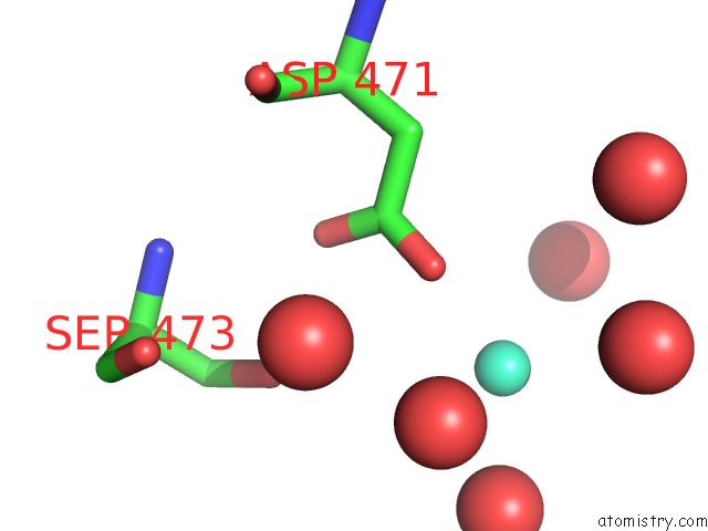
Mono view
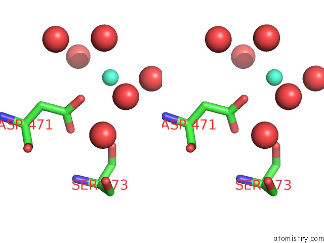
Stereo pair view
Mono view
Stereo pair view
A full contact list of Terbium with other atoms in the Tb binding
site number 2 of Crystal Structure of the Extracellular Domain of Pknh From Mycobacterium Tuberculosis within 5.0Å range:
|
Reference:
A.Cavazos,
D.M.Prigozhin,
T.Alber.
Structure of the Sensor Domain of Mycobacterium Tuberculosis Pknh Receptor Kinase Reveals A Conserved Binding Cleft. J.Mol.Biol. V. 422 488 2012.
ISSN: ISSN 0022-2836
PubMed: 22727744
DOI: 10.1016/J.JMB.2012.06.011
Page generated: Fri Oct 11 08:14:07 2024
ISSN: ISSN 0022-2836
PubMed: 22727744
DOI: 10.1016/J.JMB.2012.06.011
Last articles
Cl in 8OQ9Cl in 8OQE
Cl in 8OQG
Cl in 8OQF
Cl in 8OQA
Cl in 8OQC
Cl in 8OQD
Cl in 8OJE
Cl in 8OOD
Cl in 8OQ7